Cancer may be shaped by physical forces and not just genetic or molecular factors
Imagine being in a dense crowd. If one person suddenly starts pushing, the effect does not stay local. It spreads. People adjust, resist, or move aside. The behaviour of one individual is shaped, and often constrained, by everyone around them.
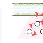
Something similar happens in our body’s tissues. Within tissues, cells are controlled by several physical constraints, like stiffness and geometry. If a cell begins to behave differently, say, because of a mutation, its neighbours immediately feel that change. These behaviours are shaped by physical cues from the cell’s surroundings.
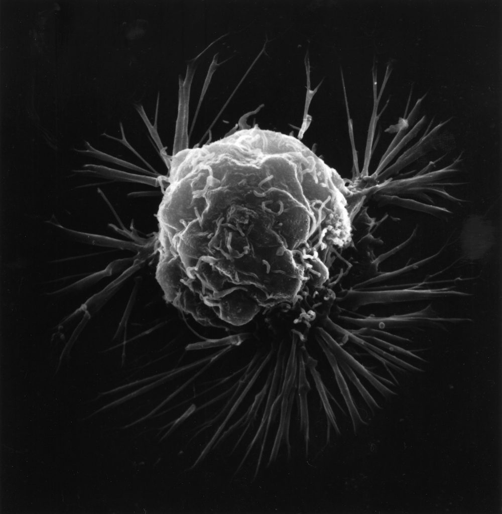
The same thing plays out in cancer cells. But researchers are now beginning to realise that some of these physical interactions with their neighbours and surroundings may be key to shaping cancer origin and spread. They are increasingly thinking of cancer as not just a genetic disease, but also a physical one.
“Cancer starts as a genetic disease,” says Medhavi Vishwakarma, Assistant Professor in the Department of Bioengineering, IISc, whose lab extensively studies the mechanics of epithelial tissues. “But what keeps cancerous cells in a tissue and what determines their fate is really an outcome of both biology and mechanics.”
Medhavi is among several researchers exploring cancer from the lens of tissue mechanics. Cancer cells, she explains, are not acting alone. “The mechanics of the cancer cells, the surrounding tissue, and even the properties of the extracellular matrix together determine how tumours grow, spread, and respond to therapy,” she says. “If we ignore that, we are essentially shooting in the dark.”
When a cell acquires a mutation, its neighbours respond. “They push back, rearrange, sometimes even try to eliminate the aberrant cell,” Medhavi explains. “So, whether a mutated cell persists is not just its own decision – it is negotiated with the tissue.”
To study this, her lab activates oncogenic pathways in a small fraction of cells within an otherwise healthy epithelial tissue and tracks how the cells respond. “We see cells around these newly transformed cells start to move differently,” she says. “There are changes in how forces are distributed, how cells rearrange, and even how pressure builds up within cells.” In some cases, healthy tissues actively expel abnormal cells – a process known as epithelial defence against cancer against cancer. But whether this succeeds depends on the mechanics of the tissue.
A recent study from her lab reported that the same oncogenic mutation can manifest differently depending on the mechanical context of the host tissue. In breast epithelium, for instance, certain mutated cells are often pushed out or constrained. In lung epithelium, the same mutation causes cells to behave more aggressively, and they tend to start colonising the surrounding healthy tissue.
“The key difference comes down to the tension at the interface between normal and transformed cells,” she says. “That determines whether the cells are eliminated, contained, or allowed to expand.”
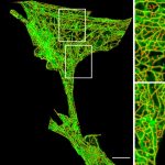
What is particularly striking is how early these changes occur. “In our experiments, these changes happen within hours,” Medhavi says. “We see things like the formation of actin structures that physically restrain cancer cells, or changes in how cells exert forces on each other.” By contrast, many biological markers typically associated with cancer either appear much later or are difficult to detect at such early stages and are still not very well understood.
“That’s something we still don’t fully understand,” she adds. “The biological markers of cancer initiation are not very well established. It would be interesting to directly compare them with the mechanical changes we observe.”
This means that if the earliest signs of cancer are mechanical rather than genetic or chemical, it might be possible to diagnose it much earlier.
Physical rules of cancer metastasis
Mechanics appears to shape not only how cancer begins, but also how it spreads.
Metastasis – the process by which cancer spreads through the body – is often described as chaotic. Cells break away from a tumour, invade surrounding tissue, and migrate unpredictably. But this view may be incomplete.
“A lot of work from the 1960s onwards focused on identifying the genes and proteins that control cell division and migration,” says Ramray Bhat, Associate Professor in the Department of Development Biology and Genetics, IISc. “That gave us what we now call the hallmarks of cancer.”
But that framework has limits.
“To understand why outcomes differ between patients, or even between cells within the same tumour, we need to look beyond genetics,” he says. “We have to think of cancer not just as a collection of cells, but as a biomaterial.” In this view, tumours behave like active physical systems – constantly deforming, interacting, and adapting under forces, much like other soft materials.
Ramray’s lab studies how cancer cells move through tissues using a combination of experiments and computational models. “What we are interested in,” he explains, “is whether cancer cells behave in ways similar to normal cells that also migrate – and whether there is an underlying logic to that behaviour.”
Cancer cells can migrate in multiple ways – as cohesive groups, streams, small clusters, or individual cells – depending on the physical properties of their environment. “If the microenvironment becomes denser or stiffer, the migration strategies change,” explains Ramray. In some cases, cells move collectively, maintaining connections with one another. In others, they disperse into their surroundings. These behaviours closely resemble processes seen during embryonic development, where groups of cells coordinate their movement to form tissues and organs.
This perspective reframes metastasis as a process shaped by physical constraints, adaptation, and collective behaviour. It also challenges a long-standing assumption – that the most aggressive cells are the ones that spread.
“Metastasis is an extremely stressful process,” Ramray notes. “Not all cells survive it. Some very aggressive ones may go extinct, while others that are more optimal – not necessarily the fastest – may succeed.”
Physics of immune surveillance
As tumours grow, they encounter another layer of complexity – the immune system. Traditionally immune responses have been understood in biochemical terms: receptors recognise molecular signals, triggering cascades that lead to the destruction of misfit cells.
But here too, mechanics might play a role.
“Immune surveillance is, at its core, a mechanical process,” says Sudha Kumari, Assistant Professor in the Department of Microbiology and Cell Biology, IISc, who studies how T cells recognise and respond to cancer cells. “These cells are constantly moving through tissues, changing shape, and physically scanning their environment to decide whether something is wrong.”
T cells, she explains, do not simply detect chemical signals. They also rely on physical cues to make decisions.
“One of the simplest examples is how the T cell receptor works,” she says. “If you present the same signal in a soluble form, the T cell doesn’t respond. But if you anchor that signal – so the cell can physically pull on it – activation happens.” This ability to “pull” on targets allows T cells to extract information that is not accessible through chemistry alone.
However, the tumour microenvironment complicates this further. “A tumour is mechanically very diverse,” Sudha notes. “The cells themselves can have different stiffness, and the environment around them keeps changing – especially during metastasis.”
These variations can affect how effectively immune cells respond. In fact, recent work suggests that tumour cells may exploit mechanics to evade immune detection.
“There seems to be a range of mechanical properties within which T cells can effectively recognise a target,” she explains. “If the tumour cells fall outside that range, the T cells may fail to respond properly.”
In other words, even if the “right” biochemical signals are present, the immune response can be muted if the physical cues are not favourable. Without these mechanical cues, T cells fail to launch the signalling cascade that leads to the release of cytotoxic molecules. This means that even if a T cell detects a cancer cell via antigen recognition, it may fail to kill it if the cell’s “mechanical stiffness” is not high enough to activate the necessary mechanisms.
This idea that mechanics can regulate immune activation is still emerging but has crucial implications in cancer-immune cell interactions. “People are now actively thinking about how to use these mechanical principles for therapy,” says Sudha. “In principle, even weak signals could be amplified if you can tune these mechanical interactions.”
Beyond stiffness, other physical properties may also play a role.
“Things like mobility, viscoelasticity, or how quickly a material relaxes after deformation can also influence how cells respond,” Sudha notes.
Taken together, these findings suggest that immune cells are not just biochemical sensors, but physical ones as well. And in the context of cancer, that distinction may matter. Because if tumour cells can alter not just their molecular signals, but also how they react physically to the immune system, they can evade the latter. At the same time, this raises the possibility of targeting these mechanical changes to design more effective immunotherapies.
But understanding these dynamics requires cell culture models that go beyond 2D Petri dishes.
Recreating tumours in the lab
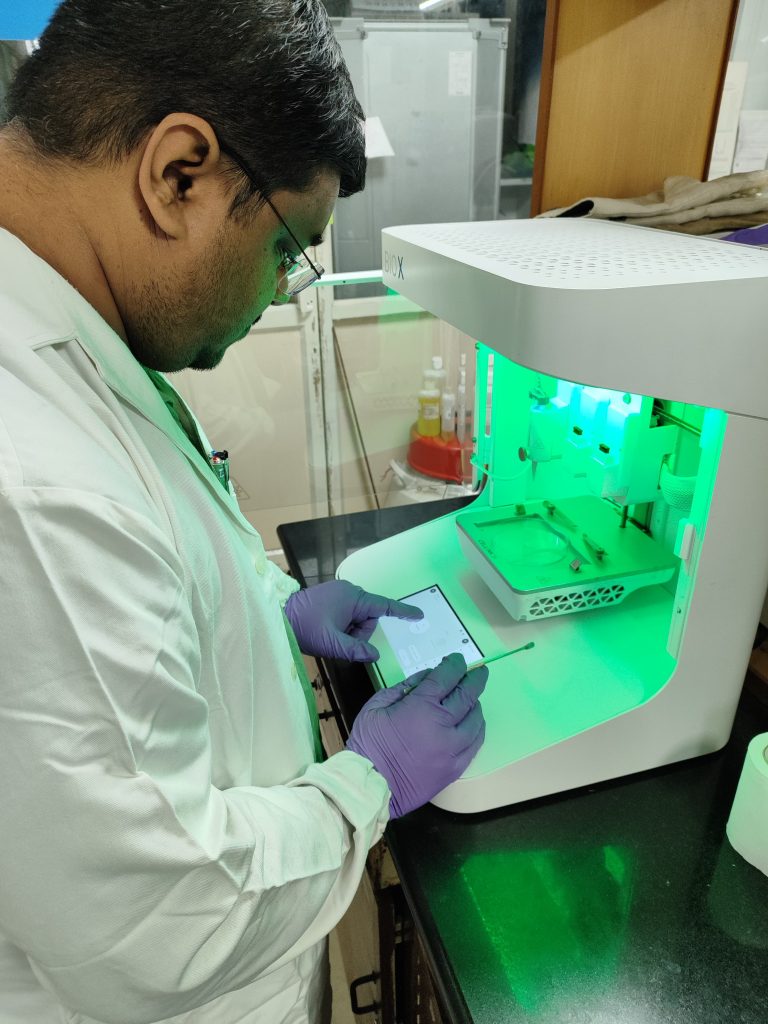
To mimic how cancer cells move in realistic environments, Kaushik Chatterjee’s lab develops 3D scaffolds that mimic the physical environment surrounding cells inside the body. These biomaterials allow scientists to precisely control properties such as stiffness, geometry, and chemical composition.
“One simple way to connect physics and cancer it to think about how we detect cancer,” says Kaushik, Professor and Chair of the Department of Bioengineering, IISc. “It often starts with a lump – a physical change in tissue.”
“If a cell needs to move through tissue, it cannot remain rigid,” he adds. “It has to deform, and it also modifies its surroundings to enable that movement.”
Traditional cell culture methods, which grow cells on flat, rigid surfaces, fail to capture this complexity. “What works in a Petri dish often doesn’t translate to patients,” he says. “A big reason is that the physical environment is completely different. When we place cells in more realistic environments, their behaviour changes,” he says. “Their growth, migration, and even drug responses are different.” This has important implications for therapy.
By engineering experimental models that replicate the complex environments found inside the body, researchers like Kaushik hope to better predict how cancers behave – and how they respond to treatment.
Ultimately, understanding cancer may require looking beyond genes to the physical rules that govern how cells coexist within tissues. Because, as Ramray puts it, cancer is not just a “collection of cells” but also a “biomaterial,” one that bends, pushes, adapts, and evolves within the body’s physical landscape.