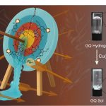
Scientists at IISc have developed a new approach to potentially detect and kill cancer cells, especially those which form a solid tumour mass. They have created hybrid nanoparticles made of gold and copper sulphide, which can kill cancer cells using heat, and enable their detection using sound waves, according to a study published in ACS Applied Nano Materials.
Early detection and treatment are key in the battle against cancer. Copper sulphide nanoparticles have previously received attention for their application in cancer diagnosis, while gold nanoparticles, which can be chemically modified to target cancer cells, have shown anticancer effects. In the current study, the IISc team decided to combine these two into hybrid nanoparticles.
“These particles have photothermal, oxidative stress, and photoacoustic properties,” says Jaya Prakash, Assistant Professor at the Department of Instrumentation and Applied Physics (IAP), IISc, and one of the corresponding authors of the paper. PhD students Madhavi Tripathi and Swathi Padmanabhan are co-first authors.
When light is shined on these hybrid nanoparticles, they absorb the light and generate heat, which can kill cancer cells. These nanoparticles also produce singlet oxygen atoms that are toxic for the cells. “We want both these mechanisms to kill the cancer cell,” Jaya Prakash explains.
The researchers say that the nanoparticles can also help diagnose certain cancers. Existing methods such as standalone CT and MRI scans require trained radiology professionals to decipher the images. The photoacoustic property of the nanoparticles allows them to absorb light and generate ultrasound waves, which can be used to detect cancer cells with high contrast once the particles reach them. The ultrasound waves generated from the particles allow for a more accurate image resolution as sound waves scatter less when they pass through tissues compared to light. Scans created from the generated ultrasound waves can also provide better clarity and can be used to measure the oxygen saturation in the tumour, boosting their detection.
“You can integrate this with existing systems of detection or treatment,” says Ashok M Raichur, Professor at the Department of Materials Engineering, and another corresponding author. For example, the nanoparticles can be triggered to produce heat by shining a light on them using an endoscope that is typically used for cancer screening.
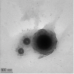
Previously developed nanoparticles have limited applications because of their large size. The IISc team used a novel reduction method to deposit tiny seeds of gold onto the copper sulphide surface. The resulting hybrid nanoparticles – less than 8 nm in size – can potentially travel inside tissues easily and reach tumours. The researchers believe that the nanoparticles’ small size would also allow them to leave the human body naturally without accumulating, although extensive studies have to be carried out to determine if they are safe to use inside the human body.
In the current study, the researchers have tested their nanoparticles on lung cancer and cervical cancer cell lines in the lab. They now plan to take the results forward for clinical development.

