Flow cytometry and microscopy are techniques routinely used in cell biology. While flow cytometry enables us to measure various biophysical parameters in cells (such as size, morphology, and internal complexity), microscopy allows us to capture high-resolution images of individual cells and the various organelles inside them.
Imaging flow cytometry (IFC) is an exciting new technique that combines both traditional flow cytometry and microscopy to simultaneously measure the biophysical parameters, and acquire an image of each cell. Conventional IFC techniques use a method called point illumination – they shine a light at a specific spot inside each cell. However, this is not useful if we need to visualise various organelles inside the cell at the same time.
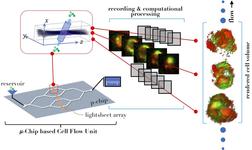
To circumvent this issue, Partha Pratim Mondal’s group at the Department of Instrumentation and Applied Physics, IISc, has developed a new system termed M3IC (Multichannel, Multi sheet and Multicolour light sheet Imaging flow Cytometry) which employs a sheet of light to illuminate the inner structures of each cell. The system consists of three major units: a multi-beam generation unit, a multi-sheet generation unit and a microfluidic chip-based cell flow unit.
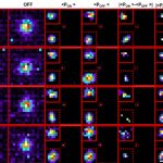
As cells flow one after the other in a channel, multiple sheets of light are lined up vertically to interrogate the cells at high resolution. The team could visualise differences in the shape and distribution of mitochondria in both normal cells as well as cancer cells treated with a chemotherapy drug. With this technique, they were able to get sharper images compared to traditional confocal microscopy, especially at low cell flow rates.
This advancement shows that now it is not only possible to count, analyse and visualise cells as a whole, but also the organelles inside them. This can be done for thousands of cells at the same time (high throughput) allowing researchers to design more efficient and cost-effective experiments.

